Human ear
The ear is the organ that detects sound. It not only receives sound, but also aids inbalance and body position. The ear is part of the auditory system.
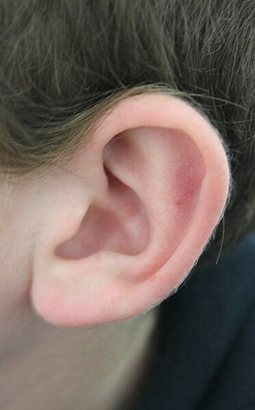
The word "ear" may be used correctly to describe the entire organ or just the visible portion. In most mammals, the visible ear is a flap of tissue that is also called the pinnaand is the first of many steps in hearing. In humans, the pinna is often called the auricle.Vertebrates have a pair of ears placed somewhat symmetrically on opposite sides of the head. This arrangement aids in the ability to localize sound sources.
Introduction to ears and hearing
Audition is the scientific name for the sense of sound. Sound is a form of energy that moves through air, water, and other matter, inwaves of pressure. Sound is the means of auditory communication, including frog calls, bird songs and spoken language. Although the ear is the vertebrate sense organ that recognizes sound, it is the brain and central nervous system that "hears". Sound waves are perceived by the brain through the firing of nerve cells in the auditory portion of the central nervous system. The ear changes sound pressure waves from the outside world into a signal of nerve impulses sent to the brain.
The outer part of the ear collects sound. That sound pressure is amplified through the middle portion of the ear and, in land animals, passed from the medium of air into a liquid medium. The change from air to liquid occurs because air surrounds the head and is contained in the ear canal and middle ear, but not in the inner ear. The inner ear is hollow, embedded in the temporal bone, the densest bone of the body. The hollow channels of the inner ear are filled with liquid, and contain a sensory epithelium that is studded with hair cells. The microscopic "hairs" of these cells are structural protein filaments that project out into the fluid. The hair cells are mechanoreceptors that release a chemical neurotransmitter when stimulated. Sound waves moving through fluid push the filaments; if the filaments bend over enough it causes the hair cells to fire. In this way sound waves are transformed into nerve impulses. Invision, the rods and cones of the retina play a similar role with light as the hair cells do with sound. The nerve impulses travel from the left and right ears through the eighth cranial nerve to both sides of the brain stem and up to the portion of the cerebral cortex dedicated to sound. This auditory part of the cerebral cortex is in the temporal lobe.
 The part of the ear that is dedicated to sensing balance and position also sends impulses through the eighth cranial nerve, the VIIIth nerve's Vestibular Portion. Those impulses are sent to the vestibular portion of the central nervous system. The human ear can generally hear sounds with frequencies between 20 Hz and 20 kHz (the audio range). Although the sensation of hearing requires an intact and functioning auditory portion of the central nervous system as well as a working ear, human deafness (extreme insensitivity to sound) most commonly occurs because of abnormalities of the inner ear, rather than the nerves or tracts of the central auditory system.
Mammalian ear
The shape of outer ear of mammals varies widely across species. However the inner workings of mammalian ears (including humans') are very similar.
The outer ear is the most external portion of the ear. The outer ear includes the pinna(also called auricle), the ear canal, and the very most superficial layer of the ear drum (also called the tympanic membrane). In humans, and almost all vertebrates, the only visible portion of the ear is the outer ear. The word "ear" may properly refer to the pinna (the flesh covered cartilage appendage on either side of the head). This portion of the ear is very vital for hearing. The outer ear does help get sound (and imposes filtering), but the ear canal is very important. Unless the canal is open, hearing will be dampened. Ear wax (cerumen) is produced by glands in the skin of the outer portion of the ear canal. This outer ear canal skin is applied to cartilage; the thinner skin of the deep canal lies on the bone of the skull. Only the thicker cerumen-producing ear canal skin has hairs. The outer ear ends at the most superficial layer of the tympanic membrane. The tympanic membrane is commonly called the ear drum. The pinna helps direct sound through the ear canal to the tympanic membrane (eardrum).
The framework of the auricle consists of a single piece of yellow fibrocartilage with a complicated relief on the anterior, concave side and a fairly smooth configuration on the posterior, convex side. The Darwinian tubercle, which is present in some people, lies in the descending part of the helix and corresponds to the true ear tip of the long-eared mammals. The lobule merely contains subcutaneous tissue.[2] In some animals with mobile pinnae (like the horse), each pinna can be aimed independently to better receive the sound. For these animals, the pinnae help localize the direction of the sound source. Human beings localize sound within the central nervous system, by comparing arrival-time differences and loudness from each ear, in brain circuits that are connected to both ears. This process is commonly referred to as EPS, or Echo Positioning System.
The complex geometry of ridges on the inner surface of some mammalian ears helps to sharply focus echolocation signals, and any sound produced by the prey. These ridges can be regarded as the acoustic equivalent of a fresnel lens, and may be seen in a large variety of unrelated animals such as the bat, aye-aye, lesser galago, bat-eared fox, mouse lemur and others.
Middle ear
The middle ear, an air-filled cavity behind the ear drum (tympanic membrane), includes the three ear bones or ossicles: the malleus (or hammer), incus (or anvil), and stapes (or stirrup). The opening of the Eustachian tube is also within the middle ear. The malleus has a long process (the manubrium, or handle) that is attached to the mobile portion of the eardrum. The incus is the bridge between the malleus and stapes. The stapes is the smallest named bone in the human body. The three bones are arranged so that movement of the tympanic membrane causes movement of the malleus, which causes movement of the incus, which causes movement of the stapes. When the stapes footplate pushes on the oval window, it causes movement of fluid within the cochlea (a portion of the inner ear).
In humans and other land animals the middle ear (like the ear canal) is normally filled with air. Unlike the open ear canal, however, the air of the middle ear is not in direct contact with the atmosphere outside the body. The Eustachian tube connects from the chamber of the middle ear to the back of the nasopharynx. The middle ear is very much like a specialized paranasal sinus, called the tympanic cavity; it, like the paranasal sinuses, is a hollow mucosa-lined cavity in the skull that is ventilated through the nose. The mastoid portion of the human temporal bone, which can be felt as a bump in the skull behind the pinna, also contains air, which is ventilated through the middle ear.
Normally, the Eustachian tube is collapsed, but it gapes open both with swallowing and with positive pressure. When taking off in an airplane, the surrounding air pressure goes from higher (on the ground) to lower (in the sky). The air in the middle ear expands as the plane gains altitude, and pushes its way into the back of the nose and mouth. On the way down, the volume of air in the middle ear shrinks, and a slight vacuum is produced. Active opening of the Eustachian tube is required to equalize the pressure between the middle ear and the surrounding atmosphere as the plane descends. The diver also experiences this change in pressure, but with greater rates of pressure change; active opening of the Eustachian tube is required more frequently as the diver goes deeper into higher pressure.
The arrangement of the tympanic membrane and ossicles works to efficiently couple the sound from the opening of the ear canal to the cochlea. There are several simple mechanisms that combine to increase the sound pressure. The first is the "hydraulic principle". The surface area of the tympanic membrane is many times that of the stapes footplate. Sound energy strikes the tympanic membrane and is concentrated to the smaller footplate. A second mechanism is the "lever principle". The dimensions of the articulating ear ossicles lead to an increase in the force applied to the stapes footplate compared with that applied to the malleus. A third mechanism channels the sound pressure to one end of the cochlea, and protects the other end from being struck by sound waves. In humans, this is called "round window protection", and will be more fully discussed in the next section.
Abnormalities such as impacted ear wax (occlusion of the external ear canal), fixed or missing ossicles, or holes in the tympanic membrane generally produce conductive hearing loss. Conductive hearing loss may also result from middle ear inflammation causing fluid build-up in the normally air-filled space. Tympanoplasty is the general name of the operation to repair the middle ear's tympanic membrane and ossicles. Grafts from muscle fascia are ordinarily used to rebuild an intact ear drum. Sometimes artificial ear bones are placed to substitute for damaged ones, or a disrupted ossicular chain is rebuilt in order to conduct sound effectively.
The inner ear includes both the organ of hearing (the cochlea) and a sense organ that is attuned to the effects of both gravity and motion (labyrinth or vestibular apparatus). The balance portion of the inner ear consists of three semicircular canals and the vestibule. The inner ear is encased in the hardest bone of the body. Within this ivory hard bone, there are fluid-filled hollows. Within the cochlea are three fluid filled spaces: the scala tympani, the scala vestibuli and the scala media. The eighth cranial nerve comes from the brain stem to enter the inner ear. When sound strikes the ear drum, the movement is transferred to the footplate of the stapes, which presses it into one of its fluid-filled ducts through the oval window of cochlea . The fluid inside this duct is moved, flowing against the receptor cells of the Organ of Corti, which fire. These stimulate the spiral ganglion, which sends information through the auditory portion of the eighth cranial nerve to the brain.
Hair cells are also the receptor cells involved in balance, although the hair cells of the auditory and vestibular systems of the ear are not identical. Vestibular hair cells are stimulated by movement of fluid in the semicircular canals and the utricle and saccule. Firing of vestibular hair cells stimulates the Vestibular portion of the eighth cranial nerve.
 It has long been known that humans, and indeed primates such as the orangutan and chimpanzee have ear muscles that are minimally developed and non-functional, yet still large enough to be easily identifiable.[14] These undeveloped muscles arevestigial structures. A muscle that cannot move the ear, for whatever reason, can no longer be said to have any biological function. This serves as evidence ofhomology between related species. In humans there is variability in these muscles, such that some people are able to move their ears in various directions, and it has been said that it may be possible for others to gain such movement by repeated trials.[14] In such primates the inability to move the ear is compensated mainly by the ability to turn the head on a horizontal plane, an ability which is not common to most monkeys—a function once provided by one structure is now replaced by another.
The outer structure of the ear also shows some vestigial features, such as the node or point on the helix of the ear known as Darwin's tubercle which is found in around 10% of the population, this feature is labelled (a) in the accompanying figure.
References
by wikipidia,
Tiada ulasan:
Catat Ulasan