Eye
The eye is the organ of vision, meaning that allows a living being to capture light and then analyze and interact with its environment.
In the animal world, there are at least forty types of visual organs called 'eyes'. This diversity raises the question of the origin of visual perception. The simplest eyes are barely able to detect the difference between light and darkness while the eyes of the most complex, as the human eye can distinguish shapes and colors.
human eye
The image formation
Any mechanism forming an image should be able to perceive differences in intensity between the different directions of incidence of light. The eye should be able to detect light, identify its direction, and establish a hierarchical relationship between the signals from different directions.
Cross section of a compound eye of dragonflies
The perception of light in the eye is done with pigments, composed of two covalently linked parts: one part protein, opsin, and a lipid moiety derived from vitamin A, the chromophore. The pigment is disposed in the photoreceptor cell membrane, and is composed of 7 transmembrane helices arranged in a circle in the membrane around the chromophore. It is the absorption of a photon by the chromophore, allowing the passage of 11-cis configuration of the chromophore to all-trans configuration, which allows the sensitivity to light. Once the pigment excited, opsin allows the activation of a G protein via one of its cytoplasmic loops, which then triggers the cellular response.
Perception of management requires focus light rays from the same direction in space on a small number of photoreceptors in the retina, which should be grouped spatially. There are many ways to combine the light rays from one direction in the animal world, emerged independently during evolution. However, we can divide the methods into three broad strategies: light rays not originating from the right direction are eliminated by shading of another structure of the eye onto the retina, the rays from one direction are curved and oriented to the same point of the retina by refraction, or rays are directed onto the photoreceptor by reflection on a concave mirror arranged behind the retina. Thus, each photoreceptor or photoreceptors group detects light from one direction.
Finally, the comparison of light intensities from the same direction in space requires an integration of electrical signals provided by the photoreceptor neurons. This integration is downstream of the retina. The signal received by the brain is never absolute, and only the difference in perceived intensity between photoreceptors is retained, and not the total level of intensity. This allows the eye to adjust to ambient light. Indeed, under conditions of high light, even a difference in intensity between two receptors appear smaller, which reduces image quality.
Optical characteristics of the eye
The eyes may be more or less successful and all have unique characteristics. The different eyes of the animal world have very different optical characteristics, often related to lifestyle of the animal. The human eye can differentiate about 8 million tones in color.
Multiple eyes of a spider (Maevia inclemens)
Sensitivity
The sensitivity of the eye is the minimum amount of light that is capable of perceiving. The sensitivity depends mainly on the size of the eye, but also on its geometry and in particular the presence of other structures ombrageantes decreasing the amount of incident light. Furthermore, the sensitivity of the eye is often modulated by the animal, for example by the presence of a diaphragm in mammals varying the amount of light admitted.
Resolution
The resolution is the smallest perceptible difference in angle between two incident rays. It therefore reflects the precision of the image that the eye is capable of forming, and the amount of detail that the eye will be able to perceive. It depends on the type of optical system for forming the image and performance. It is particularly limited by the phenomenon of diffraction of light in the case of images formed by refraction. It also depends on the number of photoreceptors: the resolution is equal to the angle between the center of two adjacent receivers. However, we observe that this is rarely the density of photoreceptors that is limiting, but more often the optical system used. This shows a very fine adjustment of the number of photoreceptors in the optical system, to minimize the loss of resolution. Finally, the resolution is often not the same throughout the retina, and peripheral parts often have a lower resolution than the center of the retina.
Diversity of eyes in the animal world
Compound eyes of a fly (Holcocephala fusca)
Eye of a viper (Vipera berus)
Eye of Squid
Cat Eyes
Detecting light
In all animals, the eyes detect light through opsins. However, the specialized nerve cells in the sensitivity to light, the photoreceptor cells are very diverse. There are two main types of photoreceptors: rhabdomériques receptors and receptors ciliates.
Receptors rhabdomériques
Rhabdomériques receptors, or rhabdoms, photoreceptor cells are characterized by the presence of membrane microvilli on the receiving carrier molecules opsins, allowing an increase of the surface of light perception. These receptors are present in all living things, but are found preferentially in protostomes. Some of these receptors have changed function during evolution, and no longer participate in the functioning of the eye, but may play a role in the synchronization of circadian rhythms, for example.
During excitation of the opsin in rhabdomériques receptors, G protein-activated in turn triggers the activation of the phosphatidylinositol membrane, and releases a second messenger, inositol triphosphate. The activation of this second messenger results in the opening of sodium channels and thus depolarization of the plasma membrane.
Forming an image
There are two main types of eyes in the animal world, each appeared many times independently during evolution. In both types, the image may be formed either by shading or by refraction or by reflection.
The simple eyes or camérulaires
The eyes have only a single chamber of photoreceptors, and oppose it in the compound eyes. The image can be formed by shading as in the nautilus, by refraction as in vertebrates or by reflection as in the scallop Jacques2.The nautilus is the only example of an animal with a single eye operated by shading. This eye, then functioning as a pinhole, then qualified eye pinhole (pinhole eye in English). It consists of a concave retina of photoreceptor cells surrounded by a layer of pigmented cells preventing the entry of the light except at a small diameter hole (pinhole) facing the retina. Thus, the rays from one direction only excite a small number of photoreceptors, which are grouped on the retina. The system allows to identify the direction of light rays and thus form an image. However, the only way to increase the resolution of the image in this system is to reduce the size of the pinhole allowing the entry of light, and therefore to reduce the amount of light admitted, that is to ie the sensitivity of the eye. The aperture size can vary from 0.4 to 2.8 mm3, which allows the nautilus to favor sensitivity or resolution within the environmental conditions.
.jpg/800px-Nautilus_pompilius_(head).jpg)
Eye of nautilus
In vertebrates and some molluscs, the image is formed by refraction through the provision of material transparent to high refractive index front of the retina. This structure helps deflect the light rays and focus all the rays from one direction over a limited area of the retina and thus form an image. This is the lens that acts as a refractive structure in fish and molluscs. The lens is generally spherical in aquatic environments. The lenses of fish and cephalopods are characterized by an increasing gradient of refractive index from the outside inwards (Mathésienne lens), which allows a correct focusing of the light rays. However, some annelids and gastropods have homogeneous lenses, and vision remains relatively unclear. The lens Mathésienne appeared independently in vertebrates and cephalopods. In terrestrial vertebrates, the lens has lost some of its power refractory, and the cornea is responsible for 2/3 of the refraction of light. Some insect larvae also have simple eyes with corneal refraction, as the larva of the beetle Cicindela.
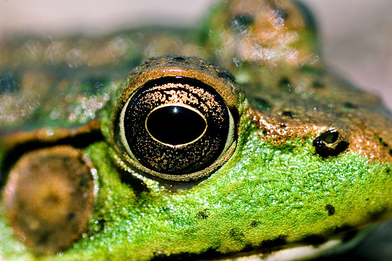
Frogeye
The eyes of the Saint-Jacques shell form an image by reflection. A concave reflective layer is placed behind the retina and acts as a mirror. Rays from one direction and are reflected differently depending on their impact on the mirror and concentrated on a small number of photoreceptors, allowing the formation of an image. There are also structures containing photosensitive mirror in some rotifers, flatworms and copepods, but the size of these structures is not sufficient to allow formation of an image.
The many eyes of this shell ajar St. Jacques are visible in the form of bright spots on the mantle edge.
The compound eyes
The compound eyes of arthropods (especially in insects and crustaceans) consist of a set of receivers (up to 30 000 in some beetles) sensitive to light which are called ommatidia. More commonly called the compound eye: compound eye. For copepods ago, in most cases, an odd look, median, which corresponds to the eye of the Nauplius larva. It is then commonly called eye nauplien.
Color perception
Some mammals such as cats or some owls are night vision. The visible spectral band varies among species. And some mammals (rats), birds (hummingbirds, swallows, pigeons ...), arthropods (lobsters, bees ...), reptiles (gecko, turtle ...) and fish (truite. ..) seem to see ultraviolet rays.
Some snakes can "see" in the infrared, but with their sensory pits. Color vision is also different depending on the species or individuals.
Are two types of photoreceptors in the retina of the human eye: rods and cones. The cones are responsible for color vision and daytime vision, they are of three types, each responsive to more red, green or blue, while the rods are responsible for the vision of light intensity and night vision. The latter being more reactive, they are used more in the reflexes and peripheral vision in the eye.
The problems of color vision, or dyschromatopsia are often grouped under the term colorblindness. The total absence of color vision is called achromatopsia.
Number and position of eyes in animals
In predators such as cats or birds of prey, the eyes are placed one beside the other which allows, in binocular vision, to better perceive the distances of prey located in front of them, in contrast, in prey such as rabbits or mice, the eyes are usually placed on either side of the head which can cover a wider field of view and to better detect the presence of a hazard in the environment.
Origin and evolution of the eye
The diversity of organisms and types of vision is, as already emphasized in Charles Darwin's Origin of Species, an intellectual challenge for the proponents of evolution. For this reason, the evolution of the eye has long been a subject of controversy between advocates of evolution and the creationists, they regard the eye as too perfect to have evolved according to the mechanisms proposed by the theory of evolution [ref. required].
There are many similarities in the functioning of the eyes of various species, such as how visual stimuli are transmitted to receivers on the central nervous system. These similarities are very numerous in amniotes. The eye of these animals derive ancestral species of the order of Captorhinidae missing there are 300 million years5.
We [who?] Was long thought that the different forms of eyes had developed independently of one species from various origins (known as development paraphyletic). However the discovery of the existence of the Pax6 gene, conserved throughout the animal kingdom and controlling eye development, has recently challenged this idea, suggesting monophyly of the eye. Now considered a primitive eye composed of a few cells developed uniquely in the animal kingdom, and afterward have diversified during the Cambrian to form at least 40 times independently of structures capable of forming images6.
Theoretical model of the evolution of the vertebrate eye.
Notes and references
by wikipidia

 The part of the ear that is dedicated to sensing balance and position also sends impulses through the eighth cranial nerve, the VIIIth nerve's Vestibular Portion. Those impulses are sent to the vestibular portion of the central nervous system. The human ear can generally hear sounds with frequencies between 20 Hz and 20 kHz (the audio range). Although the sensation of hearing requires an intact and functioning auditory portion of the central nervous system as well as a working ear, human deafness (extreme insensitivity to sound) most commonly occurs because of abnormalities of the inner ear, rather than the nerves or tracts of the central auditory system.
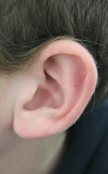
 It has long been known that humans, and indeed primates such as the orangutan and chimpanzee have ear muscles that are minimally developed and non-functional, yet still large enough to be easily identifiable.[14] These undeveloped muscles arevestigial structures. A muscle that cannot move the ear, for whatever reason, can no longer be said to have any biological function. This serves as evidence ofhomology between related species. In humans there is variability in these muscles, such that some people are able to move their ears in various directions, and it has been said that it may be possible for others to gain such movement by repeated trials.[14] In such primates the inability to move the ear is compensated mainly by the ability to turn the head on a horizontal plane, an ability which is not common to most monkeys—a function once provided by one structure is now replaced by another.

 The Mimizuka monument enshrines the mutilated noses of at least 38,000 Koreans killed during the Japanese invasions of Korea from 1592 to 1598.[7]


_(by).jpg)